1 Ibanez B, James S, Agewall S, Antunes MJ, Bucciarelli-Ducci C, Bueno H, et al. [2017 ESC Guidelines for the management of acute myocardial infarction in patients presenting with ST-segment elevation. Kardiol Pol. 2018;76(2):229–313.
2 Pendell Meyers H, Bracey A, Lee D, Lichtenheld A, Li WJ, Singer DD, et al. Accuracy of OMI ECG findings versus STEMI criteria for diagnosis of acute coronary occlusion myocardial infarction. Int J Cardiol Heart Vasc. 2021;33:100767.
3 Miranda DF, Lobo AS, Walsh B, Sandoval Y, Smith SW. New Insights Into the Use of the 12-Lead Electrocardiogram for Diagnosing Acute Myocardial Infarction in the Emergency Department. Can J Cardiol. 2018;34(2):132–45.
4 Rokos IC, French WJ, Mattu A, Nichol G, Farkouh ME, Reiffel J, Stone GW. Appropriate cardiac cath lab activation: optimizing electrocardiogram interpretation and clinical decision-making for acute ST-elevation myocardial infarction. Am Heart J. 2010;160(6):995–1003, 1003.e1–8.
5 Somers MP, Brady WJ, Perron AD, Mattu A. The prominent T wave: electrocardiographic differential diagnosis. Am J Emerg Med. 2002;20(3):243–51.
6 Nable JV, Brady W. The evolution of electrocardiographic changes in ST-segment elevation myocardial infarction. Am J Emerg Med. 2009;27(6):734–46.
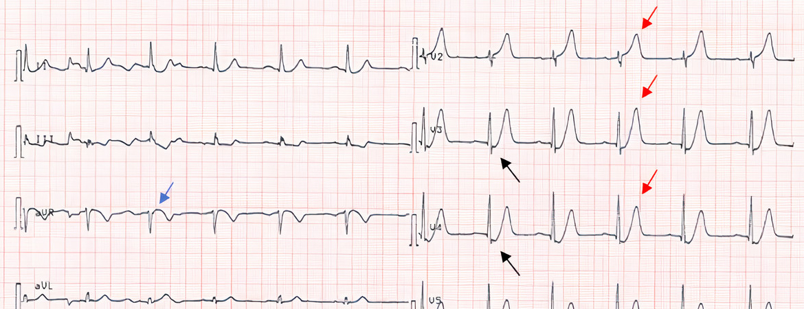
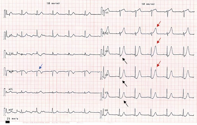
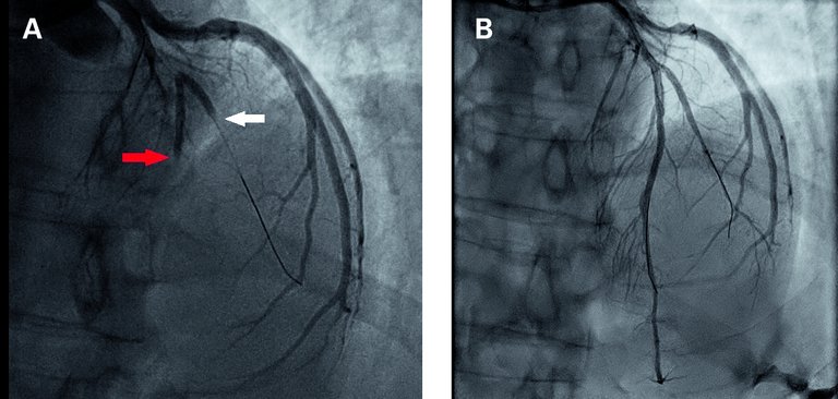
7 de Winter RJ, Verouden NJ, Wellens HJ, Wilde AA; Interventional Cardiology Group of the Academic Medical Center. A new ECG sign of proximal LAD occlusion. N Engl J Med. 2008;359(19):2071–3.
8 Verouden NJ, Koch KT, Peters RJ, Henriques JP, Baan J, van der Schaaf RJ, et al. Persistent precordial «hyperacute» T-waves signify proximal left anterior descending artery occlusion. Heart. 2009;95(20):1701–6.
9 Morris NP, Body R. The De Winter ECG pattern: morphology and accuracy for diagnosing acute coronary occlusion: systematic review. Eur J Emerg Med. 2017;24(4):236–42.
10 O’Gara PT, Kushner FG, Ascheim DD, Casey DE Jr, Chung MK, de Lemos JA, et al; American College of Cardiology Foundation; American Heart Association Task Force on Practice Guidelines; American College of Emergency Physicians; Society for Cardiovascular Angiography and Interventions. 2013 ACCF/AHA guideline for the management of ST-elevation myocardial infarction: executive summary: a report of the American College of Cardiology Foundation/American Heart Association Task Force on Practice Guidelines: developed in collaboration with the American College of Emergency Physicians and Society for Cardiovascular Angiography and Interventions. Catheter Cardiovasc Interv. 2013;82(1):E1–27.
11 Liu L, Tian J, Wang A, Yang S, Xu X, Chen Z. Clinical characteristics of patients with the de Winter electrocardiogram pattern. Zhong Nan Da Xue Xue Bao Yi Xue Ban. 2020;45(12):1431–6.
12 Montero Cabezas JM, Karalis I, Schalij MJ. De Winter Electrocardiographic Pattern Related with a Non-Left Anterior Descending Coronary Artery Occlusion. Ann Noninvasive Electrocardiol. 2016;21(5):526–8.
13 Goebel M, Bledsoe J, Orford JL, Mattu A, Brady WJ. A new ST-segment elevation myocardial infarction equivalent pattern? Prominent T wave and J-point depression in the precordial leads associated with ST-segment elevation in lead aVr. Am J Emerg Med. 2014;32(3):287.e5–8.
14 John TJ, Pecoraro A, Weich H, Joubert L, Griffiths B, Herbst P. The de Winter’s pattern revisited: a case series. Eur Heart J Case Rep. 2020;4(6):1–5.
15 Parale GP, Kulkarni PM, Khade SK, Athawale S, Vora A. Importance of reciprocal leads in acute myocardial infarction. J Assoc Physicians India. 2004;52:376–9.
16 Newman JD, Shimbo D, Baggett C, Liu X, Crow R, Abraham JM, et al.; ARIC Study Investigators. Trends in myocardial infarction rates and case fatality by anatomical location in four United States communities, 1987 to 2008 (from the Atherosclerosis Risk in Communities Study). Am J Cardiol. 2013;112(11):1714–9.